
Introduction : context and challenges
In the field of the synthesis and functionalization of inorganic nano-objects for biomedical applications, most future researches aim at developing multifunctional theranostic NPs (i.e.) that include simultaneously therapeutic and diagnostic/imaging functions allowing to envision image-guided therapy.
The projects that we develop in this research activity aim at addressing various challenges that are still to overcome in the field of theranostic magnetic nano-objects:
- The design of the dendron shell of dendronized NPs for targeting
- the design of the core of dendronized NPS : ferrite, ferrite@ferrite or ferrite@gold core-shell NPs with tunable shape and composition allowing high contrast imaging by magnetic resonance imaging (MRI), CT-scan (computed tomography-scan) and/or near-infra-red (NIR) imaging and coupled to an efficient remote therapy such as hyperthermia and/or photoablation
- the design of multifunctional organic coating and/or macromolecular vesicles bearing different functions in particular magnetic NPs and allowing furtivity, biodistribution, targeting and drug delivery,
- in vitro and in vivo validations of their efficiency.
Dendronized iron oxide NPs for MRI and displaying renal and hepatobiliary eliminations without RES captation
For biomedical applications, a key point is the design of an organic coating at ferrite NPs surface which is also challenging. Additionally to imaging, cell-targeting or drug release functions, there must be also functions preventing NPs from agglomeration in a physiological environment and favoring their biodistribution and bioelimination. The organic coating and its anchoring at the surface of NPs has to be tailored to prevent NPs’ opsonisation (the non-specific fouling of plasma protein) once entering the blood circulation and subsequent uptake by the reticuloendothelial system (RES). The final average hydrodynamic sizes of functionalized NPs has to be in the range of 10–100 nm for favoring high blood-circulation time necessary for in vivo delivery. Many synthetic and natural polymers have been used to cover the surface of iron oxide NPs however they shown various limitations (lacks of robustness, diminished impact of the core superparamagnetic properties).
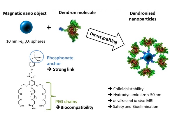
Figure1. Scheme illustrating the formation of dendronized iron oxide NPs.
As alternatives to the polymer coatings, we shown in coll. with the department of organic materials (DMO) (Dr. D. Felder) that small dendrons grafted at NPs surface were very promising as the diversity of functionalization brought by their arborescent structure simultaneously solves the problems of biocompatibility, low toxicity, high in vivo stability and specificity (Figure 1). Moreover, in addition to a controlled multi-functionalization, dendron units allow a broad versatility of size and physicochemical properties which can be precisely tuned as a function of their generation. Furthermore the grafting of dendritic molecules at the surface of 10 nm spherical iron oxide NPs using a phosphonate group as coupling agent led to a new generation of contrast agents for MRI. These nano-objects displayed relaxivity values higher than those of commercial contrast agents (polymer approach) combined to a very favorable NPs biodistribution towards tumor tissues in mices without a significant uptake in the healthy tissues (good bioelimination) (Figure 2).The use of such dendrons appears as a good way to ensure, after the grafting step, a mean particle size below 50 nm together with a narrow size distribution in suspension, both being prerequisites for a good biodistribution, i.e for avoiding RES uptake.
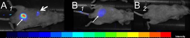
Figure 2. In vivo (A1, B1 B2) NIR fluorescence imaging of NP@dendron@fluorophore at 7 min. (A1), 20 min. (B1) and 24hours (B2) post i.v. injection. Fine arrows indicate bladder signal, large arrow indicates liver signal.
References :
– A bisphosphonate tweezers and clickable PEGylated PAMAM dendrons for the preparation of functional iron oxide nanoparticles displaying renal and hepatobiliary elimination, Chem. Commun., 2013, 49, 9158-9160.
– Effect of the nanoparticle synthesis way on dendronized iron oxides as MRI contrast agents, Dalton Transaction, 2012, 42, 2146-2157.
– Preliminary study of biodistribution and properties of MRI contrast in vivo dendrimers at the heart of iron oxide, Bulletin du Cancer, 98, 2011, S73-S74.
– Versatile dendronized iron oxides as smart nano-objects for multimodal imaging, Biomaterials, 32, 2011 8562-8573.
– Properties and suspension stability of dendronised iron oxide nanoparticles for MRI applications, Contrast Media & Molecular Imaging, 2011, 6 132–138.
– Dendronized iron oxide nanoparticles as contrast agent for MRI, Chem. Commun., 46, 2010, 985-987.
Design of dendronized NPs for targeting
Dendrons of different generations have been designed to ensure a good colloidal stability of the dendronized NPs even after coupling of targeting vectors. Dendronized NPs bearing melanin targeting ligands and a red fluorescent molecule have been injected together with dendronized NPs bearing no targeting ligand but a green fluorescent molecule. The images using confocal microscopy show a specific internalisation of NPs bearing the targeting ligands and not of the NPs without ligands (Figure 3).
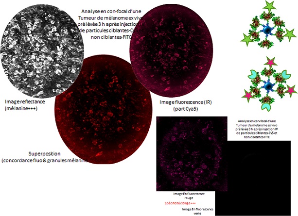
Figure 3. Confocal microscopy showing a specific internalisation of NPs bearing targeting ligands.
Design of dendronized ferrite/ferrite or ferrites/gold core shell NPs for magnetic hyperthermia (MH) and MRI
Core-shell ferrite/ferrite based NPs with different shapes and compositions are currently developed in our department for magnetic hyperthermia (MH) applications (Figure 4; synthesis). Our objective is the development of core-shell NPs with improved MH and maximized heating efficiency (SAR values) that can be optimized through the design of nanostructured core/shell ferrite/ferrite NPs with various shape, size and composition (ex: FeO@Fe3O4; Fe3O4@CoO).
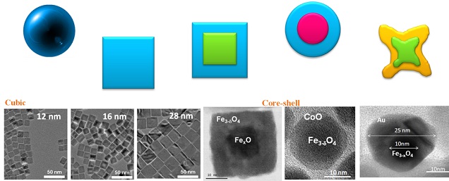
Figure 4. NPs of various shapes, sizes and core-shell structures.
In a complementary approach, we develop ferrite NPs capped with gold layer to provide core-shell ferrite-gold NPs. Such NPs are envisioned for photoablation therapy in combination with MH. Photothermal ablation is a minimally invasive therapy that typically involves photothermal sensitizers which transform the absorbed light (typically laser irradiation) into heat. NIR resonant nanomaterials such as gold permit targeted heat deposition due to their strong absorption cross section and light-to-heat conversion characteristics.
Aside the therapeutic action, imaging properties are also provided by the NPs which enable to visualize and assess the multi-therapy effects. Gold and magnetic iron oxide NPs allow respectively NIR fluorescence and MRI imaging which are highly influenced by their structural (size, shape, composition) and physical (saturation magnetization, blocking temperature…) properties at nm-scale.
Reference :
– Mastering shape and composition of dendronized iron oxide nanoparticles to tailor magnetic resonance imaging and hyperthermia, Chem. Mater., 2014, DOI: 10.1021/cm5019025.
Protein-based magnetic nanoplatforms for therapy and imaging
A novel strategy envisioned in our research activity is the encapsulation of ferrite magnetic NPs within protein-based vesicles that are well-suited to combine drug delivery with MH (and/or MRI). Human serum albumin (HSA), in addition to its intrinsic properties of biocompatibility, biodegradability and non-immunogenicity, is particularly promising to enhance NPs circulation time in the blood. A key originality of our project is the protein assembly method itself. We use a pioneering method for non-covalent macromolecular assembly based on isobutyramide (IBAM) groups (Figure 5). Such assembly approach allows the formation of original self-supported micron and submicron size particle made of proteins and also other biomacromolecules obtained by a templating method where silica (plain or porous) is used as a sacrificial template.
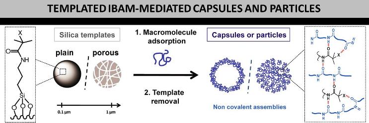
Figure 5. Scheme illustrating the formation of silica templated-assembly of submicron capsules and replicated macromolecular NPs. The macromolecules or proteins are held together via physical bridging with IBAM-graft that are hypothesized to originate from strong hydrogen-bonds.
Preliminary works demonstrated that such nano-architectures have a high potential to be used as drug delivery systems in particular for the release of various antitumor therapeutics such as doxorubicin (denoted DOX) or silencing RNA (siRNA). It was shown in coll. with the University of Melbourne and INSERM 1121 and 1110 units, Strasbourg, that DOX could be efficiently grafted at the surface of IBAM-HSA assembled microcapsules and released through protease-capsule degradation, in condition mimicking endo-lysosomes (Figure 6).
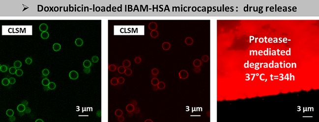
Figure 6. Confocal microscopy image of DOX-grafted IBAM-HSAFITC microcapsules from left to right, in the green channel, in the red channel, and after protease-mediated degradation.
Moreover, we recently designed multimodal protein-based nanoparticles grafted with gadolinium complexes, denoted as siRNA/HSA-Gd RNPs, which simultaneously deliver siRNA to cells and display MRI properties (Figure 7). The efficacy of such RNPs in gene silencing was demonstrated with a capacity of gene expression knockdown superior to 50% and a MRI contrast enhancement that would be suitable for visualization of targeted tumors.
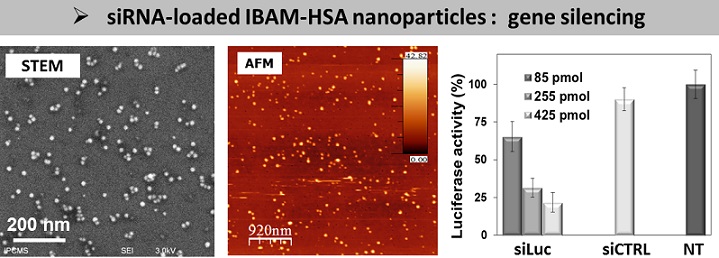
Figure 7. STEM and AFM images showing NPs made of siRNA and HSA. Gene silencing effect of the siRNA/HSA RNPs by measuring the luciferase activity of A549luc cells incubated with the same amount of RNP dispersions and having a growing dose of siRNA added per well.
References :
- Templated Assembly of Albumin-based Nanoparticles for Simultaneous Gene Silencing and Magnetic Resonance Imaging, Nanoscale, DOI: 10.1039/C4NR02623C.
- Free Standing Biopolymer-based Nanovectors, Pat. Appl. 14290121.4 (Provisional patent deposit: 25/04/2014).
- Ultrathin, Bioresponsive and Drug-functionalized Protein Capsules, Mater. Chem,. 2012, 22, 21434.
- Protein Capsules Assembled via Isobutyramide Grafts: Sequential Growth, Biofunctionalization, and Cellular Uptake, ACS Nano 2012, 6, 7584.