
Mariana Telles do Casal (Department of Chemistry, Physical Chemistry and Quantum Chemistry Division, KU, Leuven, 3001 Leuven, Belgium)
Séminaire de l’Axe 1 présenté par : Rubén Seoane Souto
Orateur: Rubén Seoane Souto (ICMM-CSIC, Madrid)
The abstract is available there.
Séminaire IPCMS présenté par : Andrey S. Klymchenko

Andrey S. Klymchenko (Laboratoire de Bioimagerie et Pathologies, UMR 7021 CNRS, Université de Strasbourg)
Abstract :
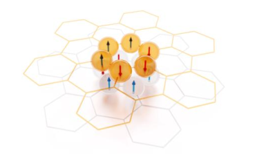
Séminaire DCMI présenté par : Elin Winkler
Elin Winkler (Magnetism and Magnetic Materials Department Bariloche Atomic Center Argentina)
Abstract
Séminaire de l’axe 1 présenté par : Olivier Simard
Speaker: Olivier Simard (Centre de Physique Théorique, École polytechnique & Collège de France)
Abstract
Séminaire Axe 3 présenté par : Prof. Rajamalli
Prof. Rajamalli (Indian Institute of Science Bangalore)
Séminaire DON présenté par : Saad Yalouz
Saad Yalouz (Laboratoire de Chimie Quantique de Strasbourg)
Résumé : Le développement des technologies liées à l’informatique quantique ouvre de nouvelles perspectives pour le calcul scientifique et la simulation de systèmes quantiques complexes. Ces avancées reposent toutefois sur une double problématique, touchant à la fois au software (logiciel) et au hardware (matériel). D’un point de vue software, nous cherchons à développer des algorithmes quantiques adaptés aux ordinateurs quantiques émergents. D’un point de vue hardware, l’enjeu est d’identifier des plateformes physiques capables de porter et de contrôler l’information quantique.
C’est précisément autour de cette double problématique software/hardware que s’articulent mes activités de recherche, à l’interface de la physique, de la chimie théorique et des sciences de l’information. Dans un premier axe software, je développe des algorithmes quantiques pour la simulation de systèmes à N-corps, avec des applications en chimie quantique et en physique moléculaire. Dans un second axe hardware, j’étudie des systèmes moléculaires complexes comme ressources potentielles pour le transport et l’encodage de l’information à l’échelle nanoscopique. Cette présentation sera l’occasion de discuter de ces deux directions de recherche et des perspectives qu’elles ouvrent pour le développement des technologies quantiques.
Séminaire des Axes 2 et 3 présenté par : Dr. Juan A. Varela
Dr. Juan A. Varela (School of Physics and Astronomy, University of St. Andrews, UK)
Séminaire Axes 4 et 1, présenté par : Christina Psaroudaki
Christina Psaroudaki (Ecole Nationale Supérieure, Paris)
Séminaire Axe 1 “Sciences et Matériaux Quantiques” présenté par : Valeria Sheina
Speaker: Valeria Sheina (Institut des NanoSciences de Paris)
The abstract is available there