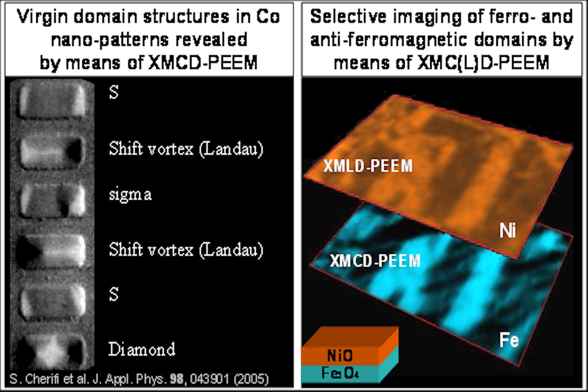
X-ray photoemission electron microscopy (PEEM) is an electron imaging technique that employs electrostatic or electromagnetic lenses to form a magnified image of the local photoelectron yield emitted from a nanostructure. The combination of PEEM with synchrotron-based x-ray photoelectron spectroscopy and the related x-ray absorption spectroscopy has allowed local spectroscopy and element selective imaging with a lateral resolution better than 25 nm. In addition to the chemical contrast, a magnetic contrast is obtained using polarized x-rays, taking advantage of circular or linear magnetic dichroism. We refer to this magnetic imaging mode as XMC(L)D-PEEM. The possibility of imaging either ferromagnetic or antiferromagnetic domains with element sensitivity makes it an advanced technique for investigating magnetic nanopatterns and multilayered magnetic films with sub-25nm lateral resolution and ps temporal resolution in the pump-probe mode. In addition to the access to the magnetic order, we have recently demonstrated the possibility of imaging ferroelectric domains in multiferroic systems using low-energy electrons in a combined PEEM-LEEM microscope.
Photoemission electron microscopy (PEEM)