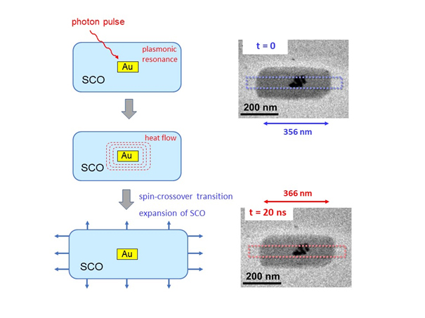
Fast photon-induced switching phenomena are the basis of nanodevices that can be operated by laser pulses. Highly promising materials for these applications are spin-crossover (SCO) compounds where a spin transition, e.g., from a diamagnetic s = 0 state to a paramagnetic s = 2 state, changes the bond length between a central metal atom and organic ligands. This transition can be induced by light or heat pulses and changes the size of the molecules. In SCO nanoparticles, reversible size changes can therefore be induced by laser pulses. While macroscopic techniques such as X-ray diffraction or magnetic measurements have already given time-resolved information on bulk ensembles of SCO nanoparticles, expansion effects at the level of individual SCO nanoparticles has so far remained difficult to study.

In a collaboration with researchers at the University of Bordeaux, ultrafast transmission electron microscopy (UTEM) at the IPCMS was applied to reveal the expansion mechanisms of individual SCO particles under nanosecond laser pulses. To facilitate the heat absorption in SCO, gold nanorods were embedded in SCO nanocrystals. Plasmonic heating of the gold rods under laser pulses leads to a controllable transfer of heat to the SCO nanoparticles which then expand rapidly due to the thermal spin transition and subsequently shrink upon cooling. With the ultrafast TEM, we are now able to measure the size of individual particles with high spatial precision and nanosecond time resolution. In this study, it is seen that elliptical SCO crystals with lengths of 200 – 500 nm expand by up to 5% within 10 – 20 ns. It is shown that the presence of plasmonic gold rods speeds up and enhances the expansion of SCO which is governed by collective effects within the nanoparticles.
Reference : Y. Hu, M. Picher, N. M. Tran, M. Palluel, L. Stoleriu, N. Daro, S. Mornet, C. Enachescu, E. Freysz, F. Banhart, G. Chastanet: Photo-Thermal Switching of Individual Plasmonically Activated Spin Crossover Nanoparticle Imaged by Ultrafast Transmission Electron Microscopy, Advanced Materials, https://doi.org/10.1002/adma.202105586


