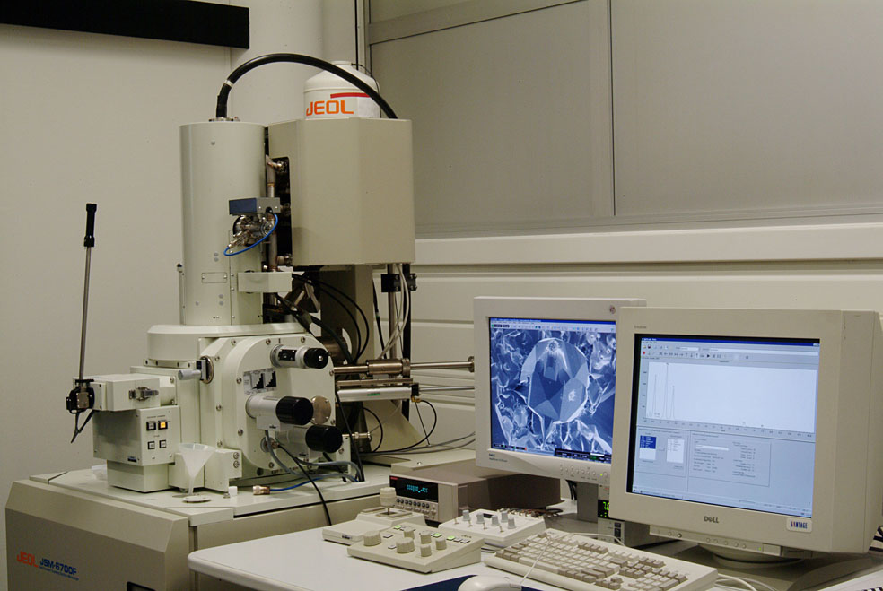
The SEM in the IPCMS through some examples
Thin film and nano-objects
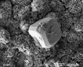
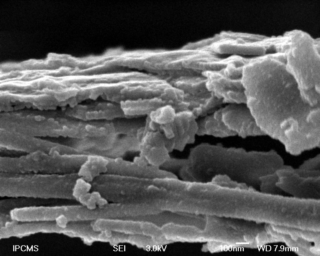
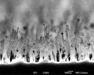
Artefacts induced by the ion beam cleaning of a Fe layer on an Ag (001) single crystal, used in the contexte of stuies of the electron spin precession in magnetic domains.
W.Weber, T. Berdot – IPCMS-DSI – SEM image J.Faerber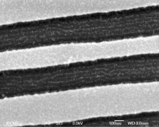
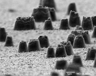
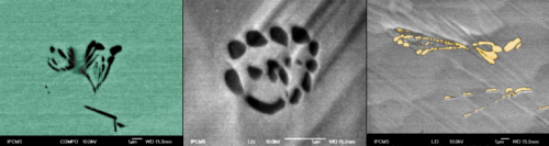
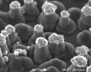
« Selective self-assembling of gold nanoparticles on PS-PMMA domains aligned in lithographied trenches. »
J-L Gallani, N. Grunsbach – IPCMS-DMO – SEM image J.Faerber
Biomaterials
A. Carrado, H. Ibrahim, J. Faerber – IPCMS-DSI – SEM image J.Faerber
A. Carrado, H. Ibrahim, J. Faerber – IPCMS-DSI – SEM image J.Faerber
Classical metallography
J-P. Kappler – IPCMS-DMONS – SEM image J. Faerber
Inorganic chemistry
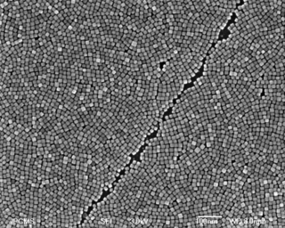
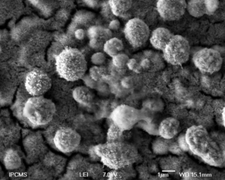
S. Bégin-Colin, B. Pichon – SEM image C. Leuvrey Iron oxide nanocubes synthesized by the thermal decomposition method and assembled in film by the Langmuir-Blodgett technique to study the magneto-transport properties.
S. Bégin-Colin, B. Pichon – SEM image C. Leuvrey
F. Roulland, A. Thomasson, M. Trassin, N Viart – SEM image C. Leuvrey
Titanium target used for pulse laser deposition to create an adhesion layer to be able to deposit platinium electrode. On left : non ablated titanium target, on right : ablated titanium target.
F. Roulland, A. Thomasson, M. Trassin, N Viart – SEM image C. Leuvrey
S. Eyele, P. Rabu, G. Rogez – SEM image C. Leuvrey
Chiral Mn III salen dicarboxylate has been intercalated in copper layered hydroxide to obtain hybrids multifunctionnal material for enantioselective catalysis.
S. Eyele, P. Rabu, G. Rogez – SEM image C. Leuvrey
Collaborations
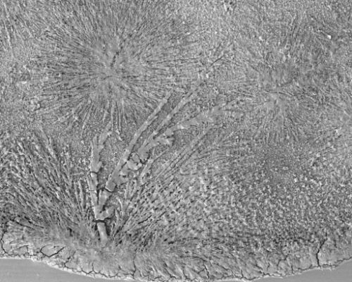
Nice geometrical craks of the carbon coating film due to some stresses in the film coming from dirt. The sample is a polymer film on a glass slide with, cold broken to see the film in cross section.
Collab. C. Arnold ICS Strasbourg – SEM image J. Faerber
Miscellaneous – Art & Science
J. Faerber, A. Carrado – SEM et OM images J. Faerber
F. Le-Normand, M. Larijani – SEM image J. Faerber
F. Le-Normand, C. Cojocaru – SEM image J. Faerber
F. Le-Normand M. Larijani – SEM image J. Faerber