B1. Introduction: what is Spin Photonics ?
Spin-photonics is a challenging research field that aims at using photons for storing and processing the information in magnetic media. Currently, the most efficient methods employed in magnetic recording make use of pulsed magnetic fields that allows writing elementary bits of information. Such approach, combined with an efficient transport of electrons without perturbing their spin states, is the core of an emerging field of science and technology named Spintronics. Alternatively, photons can be used to pattern magnetic media as shown in figure 1 and for studying the fundamental properties of the light interaction with magnetic materials. Using photons for writing and erasing elementary bits of information in magnetic media presents several advantages specific to lasers : the adjustable wavelength, the short pulses available in Ultrafast Optics, the ±h spin momentum of photons carried by circularly polarized light. Indeed these advantages are currently being used. For example the laser wavelength can be tuned to resonantly excite spin states in semiconductors; one can think of extrapolating the actual technology well above the GigaHertz bandwidth using femtosecond laser pulses; linearly or circularly polarized light allows selecting specific optical transitions in semiconductors or inducing all-optical switching in ferrimagnetic materials. Although at the present time Spin-Photonics is an emerging field that is far from outperforming the well established all magnetic technology used for example in the commercially available computer hard drives of a few hundreds Gigabytes rotating at a few thousands rd/minutes. Nevertheless, several promising achievements have been made in the past years.
Figure 1 : Spin Photonics logo patterned on a CoPt3/Al2O3 film with femtosecond optical pulses.
B2. How to study Spin Photonics ? The principles of femtosecond magneto-optical Kerr microscopy
Spin Photonics requires to investigate the magnetization dynamics on a short time scale and sub-micron spatial resolution. The femtosecond Kerr Magneto-optical microscopy is best suited for this purpose[8]. In particular we have developed a Magneto Optical Pump Probe Imaging (MOPPI) technique. In summary, it consists in a time resolving magneto-optical confocal microscope (in our case operated in reflectivity). The temporal resolution comes from two delayed laser pulses (the pump and the probe) with a duration of 150 fs and non degenerate wavelengths (800 nm for the pump and 400 nm for the probe). Focusing these beams collinearly onto the magnetic sample with a large numerical aperture microscope objective results in a spatial resolution which is diffraction limited (300 nm for our probe beam). The imaging is obtained by scanning the sample with a XY piezoelectric stage. The information on the magnetization is obtained from an ellipsometric analysis of the reflected and apertured probe beam. The quantity of interest is the normalized differential Kerr signal which in first approximation is proportional to the normalized differential magnetization ΔM/M, ΔM representing the difference between the magnetization with and without the pump pulse. We use the MOPPI technique for three different purposes : writing or erasing a pattern with a single beam, measuring the magnetization dynamics of a particular structure, and imaging magnetic structures with different levels of magnetic contrast. Figure 2 shows the spatial and temporal resolutions of our MOPPI set-up. Fig. 2a represents the magnetization curves of CoPt3 on Al2O3 (red circles) and glass (black circles) substrates. Fig. 2b shows the spatial extension of a 900 nm dot and fig. 2c a dynamical image integrated over the dot surface (inset: long delay recovery of the magnetization). In fig. 2d a spatial cross section of fig. 2b) for Y=0 is displayed.
Figure 2 : Magneto-Optical Pump Probe Imaging : spatial and temporal resolutions.
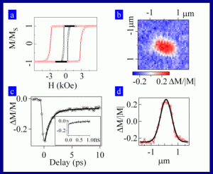
Figure 3 : Magneto-Optical writing, reading and erasing.
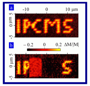
An example of magneto-optical patterning on a ferromagnetic film is displayed in figure 3. Figure 3a) represents the acronym of our laboratory IPCMS written on the CoPt3/Al2O3 sample with a pump energy density Ewrite = 8 mJcm-2 under an external magnetic field H = -50 Oe. The magneto optical pump probe imaging of the acronym is made with a density Eread = 1 mJcm-2 without external field. Applying a weak negative field allows us erasing any feature of the patterned logo by a reversed switching to the initial state. In fig. 3b), the letter M of the logo has been erased with the same pump energy Eerase = 8 mJcm-2 and H = 50 Oe. In contrast, when no external field is applied (H = 0) the effect of the pump beam is to permanently demagnetize the sample. For example, in fig. 3b) we have erased the letter C of the logo IPCMS with Eerase = 8 mJcm-2 and H = 0. The corresponding MOPPI signal is now equal to zero in that particular spatial region of the sample which is fractured into small magnetic domains.
B3. Nano-patterning of ferromagnetic films with femtosecond optical pulses
Polycrystalline ferromagnetic films are advantageous media for optical nano-patterning. We have used CoPt polycrystalline multilayers deposited on glass with a low coercive field of 370 Oe. Figures 4a) and 4b) represent two different MOPPI images of dots written respectively with the densities of pump energy Ewrite = 4 mJcm-2 (fig. 4a) and Ewrite = 8mJcm-2 (fig. 4b). In both cases there is no external magnetic field (H = 0) and the MOPPI are obtained with Eread = 1 mJcm-2. Obviously, for the lowest excitation density (fig. 4a) a dot is switched with a magnetization opposite to the environing film. In contrast, for the largest pump intensity (fig. 4b), domain structures appear. We have investigated the domain structures of these two dots by imaging them with a Magnetic Force Microscopy (MFM) apparatus. Figures 4c) and 4d) represent the corresponding MFM images of these same dots. The switched dot displays a single magnetic domain structure with an average diameter less than 500 nm. On the contrary the multi-domain fragmentation of the dot induced by the largest pump intensity clearly shows up. We could never observe the switching of a dot for H = 0 with the CoPt3/Al2O3 sample suggesting that it is the polycrystalline grain structure of CoPt/glass that allows inducing the opto-magnetic switching for H=0. The ultrafast optical control of magnetic structures without external magnetic field therefore strongly depends on the crystallinity of the magnetic material.
Figure 4 : Local optical switching and domain formation on a polycrystalline ferromagnetic film induced by femtosecond optical pulses.
