The microscope in 2020
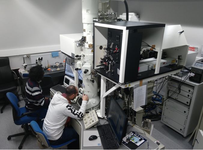
The microscope is based on a Jeol 2100 with thermionic gun that has been modified by IDES Inc. Two mirror systems allow the illumination of specimen and electron emitter with laser pulses of different wavelengths. An extra electron lens system above the condenser increases the brightness of the electron beam. The optical tables with the lasers and delay lines are coupled to the column so that the whole system is compact and supported by the anti-vibration system of the microscope. At present, a femtosecond laser (370 fs) and two nanosecond lasers (7 ns) are used. The microscope is also equipped with an electron energy-loss spectrometer (Gatan Enfinium).
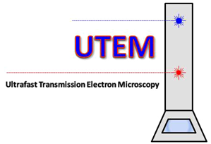
Principle of UTEM
Ultrafast Transmission Electron Microscopy (UTEM) has been developed to study microscopic objects with high temporal resolution. Conventional electron microscopy (TEM) uses continuous electron beams that hardly allow taking images of objects with exposure times below the millisecond. UTEM uses pulsed electron beams to take ‘snapshots’, so the exposure time is given by the pulse length. To achieve ultrashort and intense electron pulses, pulsed lasers are used that generate electron emission from a photocathode. The electron pulses are accelerated and focused before they traverse the specimen. The optics for imaging or diffraction is the same as in a conventional TEM.
To study the temporal evolution of nanoscale materials, a pump laser pulse of variable wavelength is used that excites the object. After an adjustable time, the electron bunch, serving as the probe pulse, traverses the object, forms the image, and is recorded by a CCD camera. Such pump-probe experiments are applied since a long time in optical spectroscopy with two pulsed laser beams or in X-ray diffraction where very short timescales are achieved but not high spatial resolution. Now, UTEM allows us to work at both high spatial and high temporal resolution. At picosecond resolution, the sub-nanometer scale is reached which is much beyond the spatial resolution of other techniques.
Operation modes of UTEM
The UTEM in Strasbourg allows the operation in the stroboscopic and single-shot mode.
1. Stroboscopic mode: reversible processes
If the specimen recovers immediately after a laser pulse, the microscope is operated in the stroboscopic mode. Here, a train of pulses is sent onto the specimen and the gun so that the same experiment is repeated many times while the shutter of the camera or spectrometer remains open. This allows working with a low number of electrons in one pulse; in the extreme case, single-electron pulses can be achieved. A femtosecond laser is used to create ultraviolet and infrared pulses at the same time. This is achieved by using an infrared laser, a beam splitter, and a nonlinear element for frequency conversion to send an ultraviolet beam onto the photocathode. The delay between pump and probe pulses is adjusted by an optical delay line. Stroboscopic experiments on longer timescales can be carried out with two electronically coupled nanosecond lasers (see setup below).

2. Single-shot mode: irreversible processes
The single-shot mode uses just one very intense electron pulse to take an exposure. This mode is applied when the specimen does not recover within a short time after the laser-induced excitation. Here, two lasers are synchronized by an electronic delay. The exposure of the image needs a large number of electrons that have to be compressed into a small pulse. Mutual repulsion of the electrons leads to broadening in space and time while the pulse propagates through the microscope. Therefore, a nanosecond laser is used for creating photoelectron pulses with a certain temporal length so that the pulses are also spatially extended in the direction of the electron beam. This reduces the mutual repulsion of the electrons. The setup of two electronically synchronized nanosecond lasers can also be operated in a stroboscopic mode at repetition rates up to 20 Hz.

Specifications of the microscope
| acceleration voltage | 80 – 200 kV |
| electron emitter | Ta disc, Ta tip, LaB6 tip photoemission or thermal emission |
| objective lens | high-resolution pole piece Lorentz config.: (B < 2 mT, magnification 40k max) |
| EELS | Gatan Enfinium |
| camera | CCD Gatan Ultrascan 2k x 2k |
| operation modes | stroboscopic, single-shot, continuous (thermal) |
| femtosecond laser | Amplitude Satsuma (370 fs, 10 W, < 40 MHz) |
| nanosecond laser 1 | Litron Nano-T 250 (7 ns, < 20 Hz) |
| nanosecond laser 2 | Quantel Q-smart 100 (7 ns, < 20 Hz) |
| delay line with fs laser | optical (fs – 3 ns) |
| delay line with ns lasers | electronic (7 ns – seconds) |
| temporal resolution with fs laser | > 1 ps |
| temporal resolution with ns lasers | 7 ns |
| spatial resolution stroboscopic | 0.23 nm (but limited by laser excitation of object) |
| spatial resolution single-shot | > 20 nm (under development) |
| EELS resolution stroboscopic | > 0.8 eV |
| EELS resolution single-shot | > 2 eV demonstrated, typ. 20 – 30 eV in quantitive core-loss EELS |
| specimen holders | double-tilt, heating, cryo, tomographic, STM (Nanofactory) environmental (gas liquid) under development |